


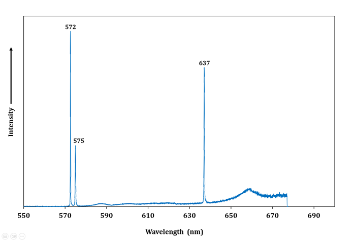
Fig.3: Raman microscope spectroscopy with N-V centres related peaks of CVD 1.547ct, emerald cut.
Home GSI Lab Note: Rare phosphorescence in CVD Lab Grown Diamond Fig.3: Raman microscope spectroscopy with N-V centres related peaks of CVD 1.547ct, emerald cut.
Fig.3: Raman microscope spectroscopy with N-V centres related peaks of CVD 1.547ct, emerald cut.
- Advertisement -


